
DC-N3 PRO
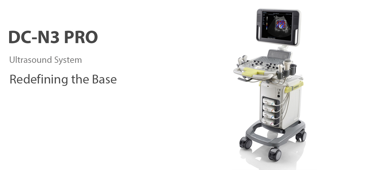
DC-N3 PRO is the answer for your requirements of high image quality, versatility and affordability. The best in class, DC-N3 PRO, is truly a redefinition of the base, providing you with much more than just an ordinary ultrasound imaging system. With advanced features and the most competitive price in the industry, it’s all about helping you raise your expectations. DC-N3 PRO is a fully featured color Doppler system that supports your needs for faster, more reliable and more accurate diagnostics. With the best in class performance, efficiency and design, you can be assured of an outstanding ultrasound experience. With its compact, user friendly and ergonomic design, it can be moved, used and positioned as per your requirements limitlessly.
Purified Harmonic Imaging for better contrast resolution providing clearer images with excellent resolution and less noise.
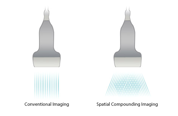
Permits use of multiple scanned angles to form a single image, resulting in enhanced contrast resolution and improved visualization.
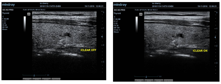
Gain improved image quality based on auto structure detection
- Sharper & Continuous Edges.
- Smooth Uniform Tissues.
- Cleaner ‘no echo areas’.
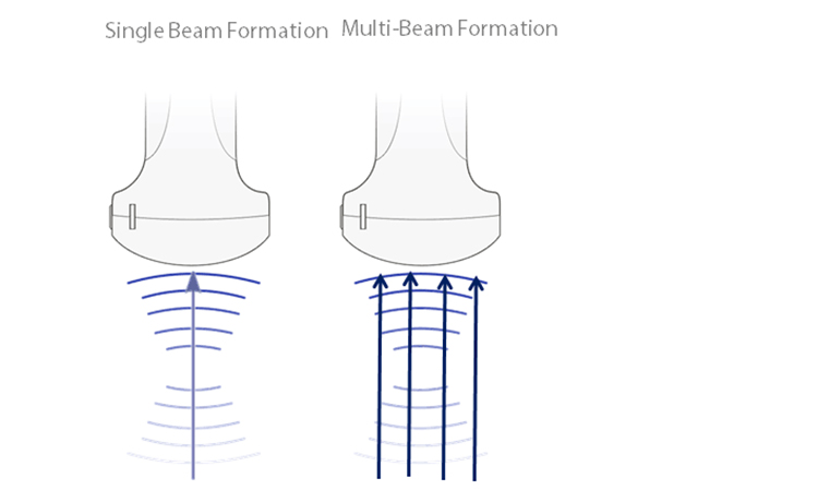
Maximum 4 times tasking for one transmitted beam, resulting in excellent time resolution and higher frame rate.
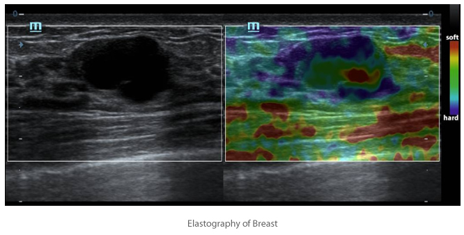
Based on Instromedix India’s latest patented technology, Natural Touch Elastography reduces dependence on user operation technique, improving operator’s reproducibility for higher clinical utility.
- Higher stiffness sensitivity.
- Good stability and reproducibility.
Get a complete and extended view of the anatomical structure through panoramic imaging coupled with velocity indication and forward/backward scan ability making scanning much easier, smoother and more controllable.
Instromedix India’s unique contrast imaging technology, utilizing contrast agent characters with both 2nd harmonic and non-linear fundamental signal to get improved S/N ratio for better diagnostic details and longer contrast agent duration for better observation.
Discover better diagnostic information through extended view of the anatomical structure on all convex and linear probes
Your tool for deeper biopsy: allowing adjustments to the scan line to gain better visibility of the needle, nerves and small vessels.
Gain precise anatomical observation by freely placing sample lines at any angle. Attain better images through simultaneous display of up to 3 sample lines.
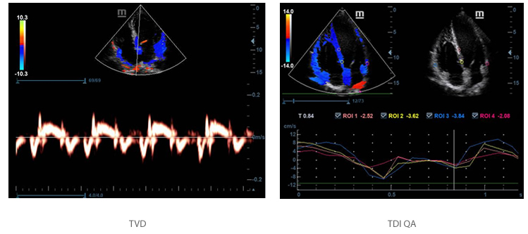
Accurately evaluate myocardial motion at different phases, and simultaneously determine myocardial synchronization. Higher frame-rate providing you with accurate results.
The Tissue Doppler Imaging (TDI) allows you to quantitatively evaluate local myocardial movement and function, with quantitative velocity parameter in TDI QA.
Dedicated inbuilt tutorial software
- Anatomical diagram illustrations including schematic and ultrasound picture.
- Standard ultrasonogram comparison with real-time scanning.
- Scanning reference picture demonstrating appropriate patient position and probe placement.
- Tips on scanning skills and diagnostic information.
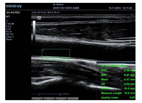
Auto measurement of anterior and posterior wall thickness providing accurate carotid status.
Simple measurement procedure for left ventricle, enhanced by auto-trace functionality and easy manual correction.
Enables optimum flexibility for post processing of the stored images including parameter adjustments, adding comments and measurements, allowing maximum productivity during scanning.
Gain instant full screen view on the click of a single key.
Gain instant auto image optimization in B, Color and PW Modes on the click of a single key.
- 180° rotatable HD monitor.
- DVD-RW drive and USB ports.
- Rotatable and height adjustable control panel.
- User-programmable keys.
- Cable management design.
- 4 active transducer sockets.
- Built-in battery supporting real-time scanning.

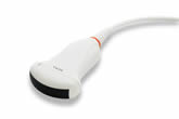
3C5A
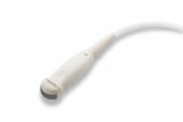
6C2

CW5s
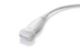
7L4B
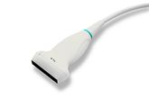
7L5
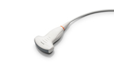
C6-2
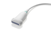
L7-3
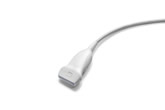
L9-3E
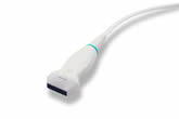
L14-6
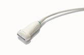
L14-6NE
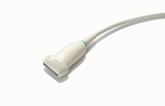
L12-3E
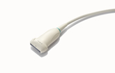
L13-3
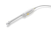
CB10-4E

