DC-8

Overview
Instromedix India’s top-of-the-range ultrasound system, DC-8 integrates the best of technology, quality and ergonomics, providing you with a unique scanning experience. Be it a fetal biometry analysis, an ovarian mass examination or showing parents the first image of their child, the DC-8 has it all. With a crystal clear image quality, comprehensive analysis package and a highly user-friendly interface, the DC-8 is your standalone solution for OB/GYN imaging.?
Performance
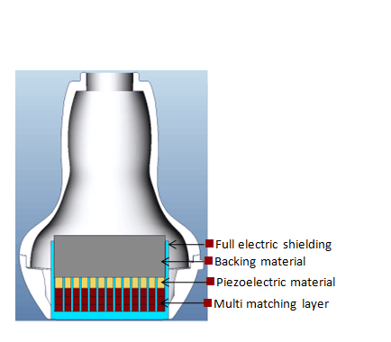
3T Transducer Technology
Instromedix India’s patent transducer technology to increase image bandwidth and transmission efficiency.
- Triple-matching layer design for higher sensitivity, wider bandwidth, and improved S/N
- Total-cut design for lower cross-talk noise, better directivity, and improved lateral resolution
- Thermal-control design for better acoustic transmission
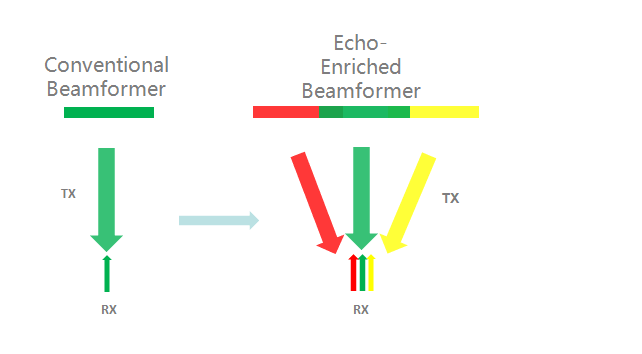
Echo-enriched Beam Forming?
Echo-enriched beam former permits the use of traditionally neglected echo signals of adjacent beams to form one finer and stronger imaging beam, providing better ‘out-of-focus’ image resolution and deeper image penetration.
PSHTM?(Phase Shift Harmonic Imaging)
Purified Harmonic Imaging for better contrast resolution providing clearer images with excellent resolution and less noise.
iBeamTM
Permits use of multiple scanned angles to form a single image, resulting in enhanced contrast resolution and improved visualization.
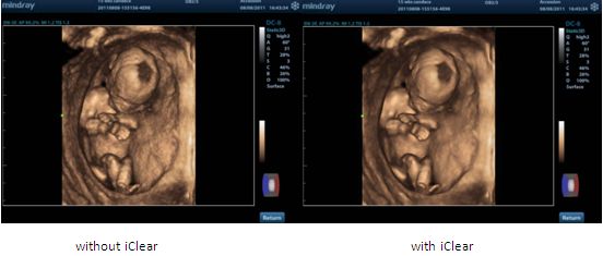
iClearTM
Gain improved image quality based on auto structure detection.
- Sharper & Continuous Edges
- Smooth Uniform Tissues
- Cleaner ‘no echo areas’
FCI (Frequency Compounding Imaging)
Permits compounding of different frequencies to form best whole field image homogeneity, providing better penetration especially for high frequency scan.
- Sharper & Continuous Edges
- Smooth Uniform Tissues
- Cleaner ‘no echo areas’
3D/4D Imaging
Including 3D/4D Flip & Sync features providing fast and easy image volume view from any direction.
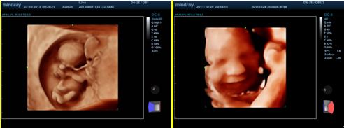
iLive
Integrating traditional ray casting algorithm with a new virtual lighting mode, iLive generates exciting real visual effects such as interactive shadowing, skin scattering and human skin-like images.
Color STIC
Spatio-Temporal Image Correlation permits advanced fetal heart diagnosis with color flow in real time 4D.
Niche
Get a 3D sectional view of the A, B and C planes to better understand the internal structure.
Curved MPRTM
Providing complete visual examination for organs and structures of different shapes by 3D volume data view in linear and curved planes.

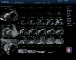
iPageTM
A CT-like view functionality displaying volume data in multiple parallel 2D images to effectively interpret anatomical structures.
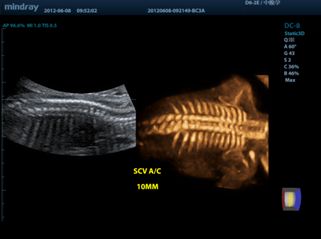
SCVTM
Slice Contrast View helps deliver higher contrast resolution and reduced noise, enabling higher-quality anatomical assessment with more information.
Workflow
Smart OBTM
Auto measurement of fetal parameters: trace and calculate the BPD, OFD, HC, AC and FL on a single click.
Smart-NT
AAuto-trace tube cavity edge with measurement result.
Smart-V
Automated trace measurements including accurate 3D VOI sketching and 3D volume size measurement.
iWorksTM
Dedicated smart tool for OB/GYN: significantly reducing the patient scan time through standardization and user-defined capability.
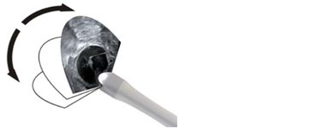
FreeViewTM
Improves patient’s comfort by automatically changing the angle of scanning plane on the Endo-cavity volume probe DE10-3E from -45° to + 45.
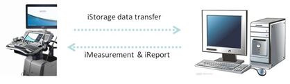
iStorageTM
Directly transfer images and reports to PC via network cable as well as convenient measurement & report template edit function.
Raw Data
Enables optimum flexibility for post processing of the stored images including parameters adjustment, adding comments and measurements, allowing maximum productivity during scanning.
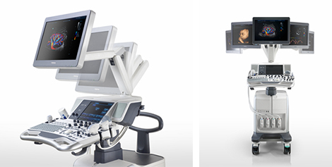
Ergonomics
- 19 inch high-resolution LCD monitor
- Articulating arm
- 10.4 inch color touch screen
- User- friendly control panel
Transducers

C5-2E
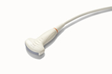
C7-3E
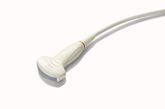
D6-2E
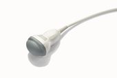
DE10-3E
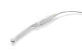
V11-3BE
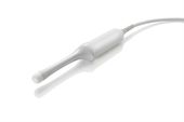
D6-2E
V11-3BE