DC-N2
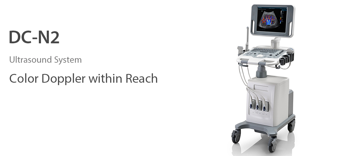
Overview
DC-N2 offers a unique combination of relevant technology, quality and affordability. Be it an experienced ultrasound professional or a beginner, the DC-N2, with its unique self-learning software and advanced imaging technologies can be used across multiple clinical settings. With a user-friendly control panel and a user-centric workflow system, the DC-N2 can be well trusted for its performance and ease of use.
Performance
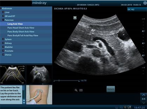
iScanHelper
- Dedicated Inbuilt Tutorial Software
- Anatomical diagram illustrations including schematic structure tips and coded tissue
- Standard ultrasonogram comparison with real-time scanning
- Scanning reference picture demonstrating adequate patient position and probe placement tips on scanning skills and diagnosis information
PSHTM(Phase Shift Harmonic Imaging)
Purified Harmonic Imaging for better contrast resolution providing clearer images with excellent resolution and less noise.
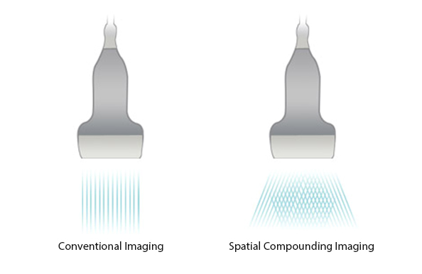
iBeamTM
Permits use of multiple scanned angles to form a single image, resulting in enhanced contrast resolution and improved visualization.
iClearTM
Gain improved image quality based on auto structure detection
- Sharper & Continuous Edges.
- Smooth Uniform Tissues.
- Cleaner ‘no echo areas’.
Multiple Beam Formation
Maximum 4 times tasking for one transmitted beam, resulting in excellent time resolution and higher frame rate.

iScapeTM
Get a complete and extended view of the anatomical structure through panoramic imaging coupled with velocity indication and forward/backward scan ability making scanning much easier, smoother and more controllable.

ExFOV
Discover better diagnostic information through extended view of the anatomical structure on all convex and linear probes
B-SteerTM
Your tool for deeper biopsy: allowing adjustments to the scan line to gain better visibility of the needle, nerves and small vessels.
Smart 3D
Smart 3D providing easy image volume view from any direction.
Workflow
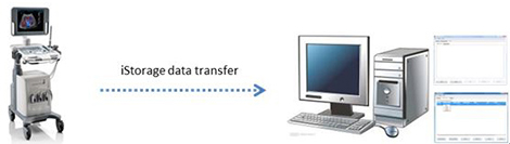
iStorage
Directly transfer images and reports to PC via network cable.
Auto IMT (Intima-Media Thickness)
Auto measurement of anterior and posterior wall thickness providing accurate carotid status.
iTouchTM
Gain instant auto image optimization in B, Color and PW Modes on the click of a single ke.
iZoomTM
Gain instant full screen view on the click of a single key.
iStationTM
Instromedix India’s unique Patient Information Management System allowing you to integrate, review, archive and retrieve patient data effectively.
Ergonomics
- 15''/17’’ LED monitor with easy handle
- Height adjustable control panel
- CD/DVD-RW and USB ports
- Removable transducer holder
- Built-in battery for live scanning
- Three active transducer connectors

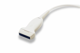
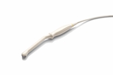
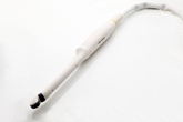
Transducers

35C50EA
Center Frequency: 3.5MHz

35C20EA
Center Frequency: 3.5MHz

65C15EA
Center Frequency: 6.5MHz
75L38EA
Center Frequency: 7.5MHz

10L24EA
Center Frequency: 10MHz

65EC10EA
Center Frequency: 6.5MHz
65EC10ED
Center Frequency: 6.5MHz
65EB10EA
Center Frequency: 6.5MHz